MRI vs. CT Scan vs. X-Ray: Which is Right for You?

Deciding between an MRI, CT scan, or X-ray can be confusing, especially when you’re not sure which is the most appropriate for your specific health concern. This guide demystifies the process, helping you understand when each type of imaging is recommended and what to consider before undergoing a scan.
Understanding Your Options
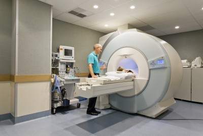
MRI (Magnetic Resonance Imaging):
Utilizes magnets and radio waves to create detailed images of the body. Ideal for diagnosing issues related to soft tissues, discs, and the central nervous system, MRIs are more sensitive but less specific, revealing details that may not always correlate with symptoms.
CT Scan (Computed Tomography):
Combines multiple X-ray images to produce a comprehensive view of your body’s interior. Best for viewing bone injuries, diagnosing lung and chest problems, and detecting cancers. However, CT scans expose you to a higher dose of radiation compared to standard X-rays.
See Also: 4 Best Exercises For People With Sciatica
X-ray:
The quickest and most accessible form of imaging, perfect for examining fractures, infections, or abnormalities in the lungs. While X-rays are less detailed than MRIs and CT scans, they are invaluable for diagnosing specific conditions.
When to Choose Each Imaging Type
- MRI: Preferred for detailed images of soft tissues, spinal conditions, and joint problems. Essential for conditions like disc herniations, where soft tissue visualization is crucial.
- CT Scan: Optimal for quickly examining injuries or conditions that require a broader view of bone and organ systems. It’s particularly useful in emergencies.
- X-ray: Often the first step in diagnosing bone fractures or lung issues. It’s the go-to option for its speed and efficiency in revealing structural abnormalities.
Before Opting Imaging Tests, Ask Yourself Three Questions:
#1 Will the imaging pinpoint the cause of my pain?
The Paradox of MRIs and CT Scans in Diagnosing Pain
Have you ever wondered if an MRI or CT scan can pinpoint exactly where your pain originates? It’s a common question with a surprising answer: often, the answer is no. Here’s why understanding the capabilities and limitations of these powerful diagnostic tools is crucial.
The Limitations of Imaging
Misleading Results: Research shows that MRIs can reveal disc bulges and other spinal abnormalities in people without any pain—15% in 15-year-olds, 30% in 30-year-olds, and 60% in 60-year-olds. These findings challenge the assumption that visible issues on an MRI or CT scan always correlate with pain.
Sensitivity vs. Specificity: While MRIs are highly sensitive, detecting minute changes within the body, they are not always specific to the cause of pain. Dr. Christopher DiGiovanni notes that it’s rare to see a completely “normal” MRI, suggesting that abnormalities are common, even in asymptomatic individuals.
The Role of Pressure: Discs can change shape under different conditions, which means lying down during an MRI will not show the pressure that causes pain when sitting or standing. This discrepancy can lead to misinterpretations, where a patient’s symptoms don’t align with imaging results.
The Importance of Clinical Diagnosis
Beyond Imaging: A thorough history and physical examination are pivotal in making an accurate diagnosis. Sometimes, imaging findings may not reflect the patient’s experience of pain, emphasizing the need for a comprehensive evaluation by a healthcare provider.
Open MRI as a Solution: Open MRI machines, which allow for scanning in various positions, can sometimes offer a clearer picture of what happens to discs under different types of pressure. However, their availability and cost can be limiting factors.
Making Informed Decisions: MRI, CT, X-ray
Despite these challenges, MRIs and CT scans play a vital role in the diagnostic process, especially when differentiating between conditions like tumours and back pain. The key is to use them judiciously, informed by a thorough clinical assessment.
Remember, if you’re facing decisions about diagnostic imaging, discussing all aspects with your healthcare provider is crucial. Understanding the strengths and limitations of MRIs and CT scans can help you make informed choices about your healthcare journey.
#2 How will I know what is causing my pain then?
Unraveling the Mystery of Your Pain: Beyond Imaging
When you’re in pain, pinpointing the exact cause can feel like searching for a needle in a haystack. It’s natural to wonder if an MRI, CT scan, or X-ray can offer quick answers. However, the journey to understanding and addressing your pain often begins long before any images are taken.
The Central Role of Clinical Evaluation:
In-Depth Consultation: Your chiropractor or physiotherapist should start with a detailed conversation and physical examination, dedicating at least 15 to 30 minutes to understanding your symptoms, lifestyle, and any potential injury mechanisms. This initial step is crucial in forming a comprehensive view of your health status.
Diagnostic Expertise: Contrary to common belief, the true art of diagnosis lies not within the images produced by an MRI or X-ray but in the skilled interpretation of your history and physical examination findings. The overreliance on medical imaging has led to a decline in traditional diagnostic skills among some healthcare practitioners.
Formulating a Diagnosis: A proficient healthcare provider will consider your clinical presentation and may already have a primary diagnosis in mind, along with a few alternatives, based on your initial evaluation. The purpose of subsequent imaging tests like MRIs or X-rays is to confirm these suspicions rather than to discover them.
SEE ALSO: 6 Things You Should Look For In A Chiropractic Clinic: Chiropractic Clinic Best Practices
#3 Choosing the Right Diagnostic Imaging: MRI, CT Scan, or X-ray?
Deciding whether an MRI, CT scan, or X-ray is the best option for diagnosing your condition depends largely on your specific symptoms and the nature of your ailment. Understanding the strengths and applications of each imaging technique can guide you and your healthcare provider toward the most effective diagnostic approach.
When to Consider Each Imaging Option: MRI, CT Scan or X-ray
X-ray for Osteoarthritis:
- Ideal for Initial Diagnosis: X-rays are often sufficient for diagnosing osteoarthritis, characterized by morning stiffness that improves after movement.
- Cost-effective: Compared to MRI and CT scans, X-rays are more affordable and widely accessible for assessing joint wear and tear.
MRI for Disc Herniations:
- Superior Soft Tissue Imaging: MRIs excel in detailing soft tissue conditions, making them perfect for identifying disc herniations.
- Non-invasive Diagnosis: Before opting for an MRI, consider chiropractic care, extension exercises or Yoga Cobra, which can effectively treat many cases of disc herniation.
See Also: The Best Exercises For Your Herniated Disc
MRI for ACL Tears:
- High Sensitivity: MRIs provide detailed images of soft tissues, including ligaments, making them more sensitive for detecting ACL tears than physical examinations alone.
- Comprehensive Damage Assessment: An MRI can also reveal associated cartilage and ligament injuries, offering a complete picture of the knee’s condition.
CT Scan for Fractures Invisible on X-rays:
- Enhanced Bone Imaging: CT scans are superior for visualizing bone fractures that X-rays may miss, such as rib fractures.
- Critical for Complex Diagnoses: In cases where a detailed bone structure view is necessary, CT scans offer the clarity needed for accurate diagnosis.
Arthrograms for Cartilage Tears:
- Specialized Imaging: Injecting dye into a joint before taking an X-ray, MRI, or CT scan can highlight cartilage tears more clearly, essential for diagnosing meniscus, hip labral, and shoulder labral tears.
Key Considerations When Choosing Between An MRI, CT and X-ray
- Symptom-Specific Imaging: The choice between MRI, CT scan, and X-ray should be guided by the specific symptoms and the area of the body affected.
- Consult with Your Healthcare Provider: A thorough clinical evaluation is crucial in determining the most appropriate imaging technique for your condition.
- Cost vs. Benefit: Consider the cost and diagnostic value of each imaging option, especially when dealing with conditions that might be managed with less expensive alternatives.
Dangers of an MRI
An MRI machine has a powerful magnet that attracts most metallic objects. Therefore the danger lies with any implants that contain iron. Your implant can move and injure you physically or heat up and even burn you. The following are implants that are unsafe for MRIs.
- Older vascular stents
- Cochlear implants
- Brain-aneurysm clips
- Pacemakers or cardiac defibrillators
- Gastrointestinal cips
- Certain medication pumps (such as insulin pumps)
- Stainless steel spinal fusion screws and plates cause problems for the MRI and you. However, titanium screws and plates are fine. The stainless steel causes the digital picture to be degraded.
- Metallic objects like metal filings that have accidentally gone into the eye.
Gadolinium dyes are used in some MRIs, Gadolinium contrast dye is safe for most of you, but unsafe and can cause death in those with bad kidneys. If you are getting a procedure with Gadolinium your medical doctor should do a screening test for your kidney function.
Understanding the Radiation Risks: CT Scans, X-rays, and the Safety of MRIs
When it comes to medical imaging, understanding the risks and benefits of each option is crucial. While CT scans and X-rays are invaluable tools in diagnosing various conditions, it’s important to be aware of the radiation exposure involved. MRI scans, on the other hand, offer a radiation-free alternative for many diagnostic needs. Let’s dive deeper into the implications of choosing between these imaging techniques.
Radiation Exposure: A Comparative Look
Everyday Radiation vs. Medical Imaging
- On average, we’re exposed to about 2 millisieverts (mSv) of radiation annually from natural sources like rocks, the sun, and even aeroplane flights.
- Residents in high-altitude areas, such as Colorado, receive around 10 mSv due to less atmospheric protection.
CT Scans and X-rays: How Much Radiation Do You Get?
- A neck CT scan exposes you to about 6 mSv, roughly three years’ worth of natural radiation.
- A brain CT scan equals about 2 mSv, or one year’s worth of background radiation.
- A low back X-ray comes in at 1.5 mSv, about eight months’ worth of natural exposure.
- Neck X-rays and mammograms offer lower exposures, at 0.2 mSv and 0.8 mSv respectively, but these still add up over time.
Assessing the Need for Imaging
Before opting for a CT scan or X-ray, consider the following:
- Diagnosis Clarity: If your diagnosis is uncertain, imaging may be crucial.
- Treatment Impact: Will the scan results change your treatment approach? If not, reassess its necessity.
- Previous Scans: Remember, radiation exposure is cumulative. Evaluate the necessity of additional CT scans carefully.
- Cancer Risk: Those at high risk or with a family history of cancer should weigh the benefits and risks more carefully.
- Pain Localization: Can the scan accurately identify the source of your pain?
MRI: A Safer Alternative?
MRIs stand out as they do not use ionizing radiation, making them a safer choice in many scenarios. Despite advances in medical technology, it’s essential to consider the historical context—lessons learned from past practices, like the widespread use of X-rays in pregnant women during the 1960s, underscore the importance of caution and informed decision-making in medical imaging.
Making an Informed Choice: MRI, CAT scan and X-ray
Choosing between an MRI, CT scan, or X-ray involves a careful evaluation of your specific situation, guided by the expertise of your healthcare provider. By understanding the strengths and limitations of each imaging method, you can make informed decisions about your health care.
Tell us what you think in the comments below and like us on Facebook. This Toronto Downtown Chiropractor will answer all questions in the comments section.






i was in Hawaii and I fell twice when taking a nature hike in the forest. I fell on my right side on some rocks. I had an x ray done but it did not show anything broken. but i get throbbing pain at times in my sidearm muscle and lifting my arm at times hurts. I can also feel and hear snapping or crunching or grinding sounds at time when I lift my are and twist it. . It feels great when I have hot water on it in the shower but after that not so well. I cant sleep on my right side because of my it and it get pretty uncomfortable at nite. I cant even do my push ups. I feel that I need a MRI because I feel that I have a tear somewhere in a ligament that the x ray did not find. What do you think.? Thank you
Author
Thanks for your question Ken. You can definitely injure soft tissue when you fall on rocks. You can cause a contusion or bruise your bone which can take up to 6 weeks to heal. Other tissues like muscles, tendons and ligaments can become stretch or even tear. It’s best to get the opinion of your chiropractor, physiotherapist or medical doctor first before deciding if advanced imaging is needed. Often a plan of management is determined and a course of treatment is laid out by your chiropractor or physiotherapist. Medical doctors can give you medication or refer you. Their job is not to treat but to diagnose you with or without the help of imaging.
While it is not common it is possible there may still be inflammation there still as you feel better temporarily but worse after your shower. If there is inflammation adding ice to the area for 10 minutes at time would be helpful. Make sure you apply a cloth next to the skin then the ice pack to protect the skin. You should never put ice directly on the skin. The above assumes there is no open would. If there is then you would need proper wound care from a medical doctor, nurse or nurse practitioner.
In summary, you should ask a health care practitioner in person if you need an MRI. Most of the time they are not necessary.
The above is an opinion and not a recommendation. Hope that helps your pain.
Hello, for the past two days I’ve had these excruciating headaches. It’s more of a feeling than a pain in my head, feels like there’s a huge pressure inside the middle. Also, when I walk I feel like I’m going to lose my balance but don’t. The room isn’t spinning, there’s no blurred vision, no slurred speech, I can remember everything about myself and my birthday. There’s no pain anywhere else on my body.
I just have this weird head pain and feeling unbalanced when I walk. I also feel nauseous but don’t throw up. I went to urgent care but they couldn’t really tell me anything, just said I might have a migraine or tension headache. My boyfriends has suggested dehydration.
I plan on going to a real hospital because I want this to go away. But do you have any idea what this sounds like?
Author
Thanks for your question Ashlyn. Your symptoms can be related to a number of things including your inner ear, the back of your brain that controls balance, arteries that go up to your brain from your neck and lastly upper cervical problems. I can’t tell you which one as I have obviously not examined you. The above is an opinion based on the limited amount of information. I don’t recommend you keep looking up websites to determine your diagnosis as often you will think you will usually think the worst. I would go to another doctor like neurologist who can differentiate your symptoms and signs and determine your likely diagnosis before getting imaging. The doctor should spend time with you getting your history and exam.
Imaging should only be done to confirm a diagnosis or to rule out some nasty problems. Ask your doctor what he/she is thinking of before getting imaging. If they have no idea either your problem is very uncommon or they are not doing a good job. Most doctors are really only confirming a diagnosis.
Hope that helps your headaches? If you have any more questions for this Toronto downtown chiropractor I will do my best to give you a useful answer.
I have neck pain since two month…not improving… Its study .pain….two xray and one MRI showing nothing…. Worry about neck pain …could its due to cancer …like head and neck cancer or spinal cancer metastatic…..
Author
Thanks for your question Anil. First, while it’s not likely you have cancer just because you have pain for two months. Cancer is excluded with a proper history. The most important questions being.
1.If you have previous cancer. Doesn’t sound like you do, otherwise, you would have mentioned it. Correct me if I am wrong.
2. Are you over 65 year of age? Again your writing doesn’t indicate this, but you can correct me.
3. Weight loss for no reason
4. Night pain so much so that you don’t sleep. You would have complained about this.
If you didn’t answer yes to these questions the chances are you don’t have cancer. Just because you don’t have anything on MRI and X-rays doesn’t mean you have cancer. Why don’t you go to a doctor that will do a thorough history rather than someone that charges you for an MRI and X-ray. Likely a good chiropractor can help.
This is an opinion and not a recommendation.
Hope that helps your spine.
Thanks ..for reply….yes I m 32….not having previous cancer….pain is light or mild….other than pain I have constant phelgm in my throat…..alrwdy visited 4 ent doc…but they think its due to post nasal dip… And gerd…
.
I strongly believe.. My neck pain is due to…throat problem……already.. Taken 5 day IFT swt .
But no use….pain is light/mild
.
But its constant
Author
Anil, if your neck pain is related to your throat pain then I cannot help as that it not my area that I deal with. I don’t deal with GERD or reflux. Sorry, I can’t help you.
Also, while you may be frustrated at not knowing what the problem is, try not to think the worse of your situation.
Dr ken; i have chronic back pain mostly left side,it goes from hip area to lower shoulder blade,have had xrays,mri,ct scan,all which have shown nothing,have seen several docs,including rhumatologist,and pain doctor. 6mths of chiro,6mths physio,massage ,accupuncture. nothing has helped,chiro and physio both seem to make it worse. if i stand more than 10 min or sit more than 10min it starts to burn and then gets to the point where its hard to breathe and i feel like i am going to pass out.if i do the physio excersises or get chiro adjustment i am in extreme pain for about a week after. they have prescribed me naproxen,cyclobenzaprine,amitriptyline,i got no relief from any of it,i have also received multiple trigger point injections,with no success. the only relief i have found is lying on my back with heating pad and pulling my knees to my chest,however as soon as i stop i am back to square one. this all started when i was 40yrs old ,am now 52. I live in saskatoon saskatchewan. the doctors around here say there is nothing more they can do for me. I use to enjoy all kinds of physical activities,now i cant do anything without extreme pain,it has ruined my life. any advice would be very helpful. thank you
Author
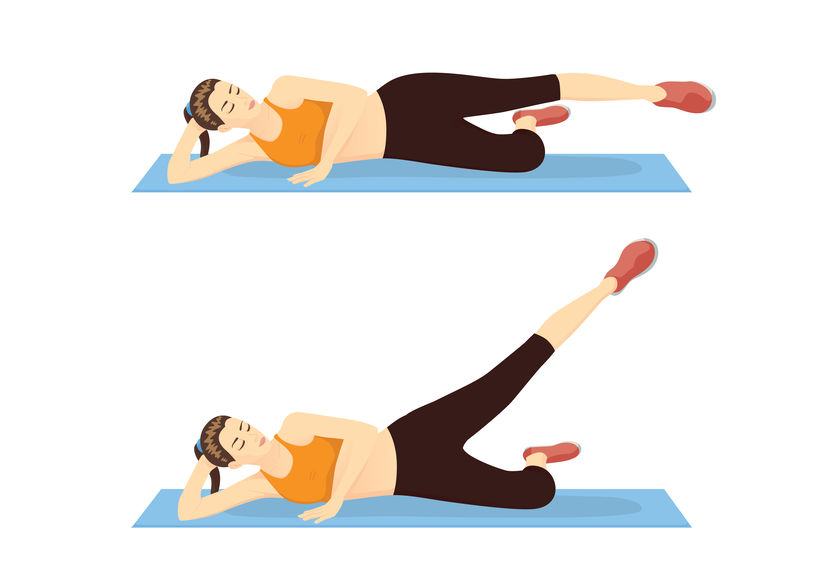
Thanks for your question Mike. Try reaching towards your feet while sitting. It’s like the knee to chest exercises but you are doing it while sitting.
http://gayyoga.gn.apc.org/Article%20part%203_files/image004.jpg
Like any exercise, it may make you worse but from what you are telling me it has a chance of making you better. You know you are getting worse if the pain increases or becomes sharp or goes further down the leg. If that happens then you should stop right away.
This is an opinion and not a recommendation.
Hope that helps your chronic lower back pain.
Hello, I had a cervical fusion c5 c6 on Nov 17 2015.. Im still suffering from neck pain and tingling numbness to right arm and severe pain to left side of neck.. Mri shows bulging disc on top and bottom of where fusion was done and xrays show it fused… Supposedly spine specialist says I have a great spine no other surgery required. My EMG shows nerve irritations and impinging…My lower back has impinging also. Nothing shows for surgery to be done im suffering from this pain Neck and Lower back. I also have weakness to my right side of my arm and I dont kno what Else to do. Plz Help.
Author
Thanks for your question Gigi. Unfortunately when you have fusion surgery what they don’t tell according to many patients that have had fusion surgery, is that you can have a problem above and below vertebrae that were fused. This includes disc herniations and pinched nerves. As far as I can tell this is an inevitable part of fusion surgery but it does not mean you will have symptoms. Research states that this take more than 10 years for really visible effects.
Hopefully this fact was disclosed to you before the surgery. I am guessing though that your pain must have been severe enough to warrant surgery and that you had tried conservative treatment such as chiropractic or physiotherapy. Surgery should always be the last resort after injections are also tried.
Also while, the surgeon says there is no need for surgery there is clear indications that the nerve is being pinched or irritated on EMG. This means that you do have a problem with the nerve that showing up objectively. As you have a fusion I am not going to give you an opinion on the type of exercises to do. However I would go find the best chiropractor or physiotherapist in your area to get some treatment done.
Please remember this is an opinion and not a recommendation.
Hope that helps your neck and arm pain with tingling.
I need help. I had a bad chiro adjustment and it sent me straight to the ER. It was fill-in never talked to them or said two words. They applied severe sudden direct pressure to my back three times and nothing cracked. Cracked my neck and I’ve never been the same. This was 8 mos ago. I went to ER and nothing showed on xrays. I for SURE thought I broke a rib and had difficulty breathing. Tried so incredibly hard to tough it out. Sharp pain when I sneezed or picked something light up in bottom of rt shoulder blade. I have had numerous xrays, mris, been to so many drs. my dx are winging of scapula, scapular dykinesis, pain in thoracic spine, herniation of cervical intervertebral disc with radiculopathy and pain of cervical facet joint. I had three steroid epidural injections, two facet injections. I am a young female. I can lift a gallon of milk but then it flares up the pain to agony. so I cannot lift or pull things with my right arm. I drive with my left and honestly I am stuck at home now 24/7 and no social life because the pain is so bad even after 8 mos. The only option Drs. are giving me is nerve ablation but I still did not get significant relief from two facet injections maybe 30-40% relief and am told the nerve ablation will only give 10-20% for 6 mos-2 years. Facet injections only worked for about a week.. sore for a week and then a little improvement but right back to intense pain. I am forced to take pain pills otherwise I physically and mentally cannot handle the pain. Is there any way there is a muscle or tendon or ligament that is torn right on the bottom of my rt shoulder blade that wouldn’t show up on mri or xray? It literally feels like I have a knife stuck in my lower right shoulder blade. I have been having muscle spasms for 8 mos now and I am guessing that other areas are having to compensate for something that is injured and unable to heal itself. Any or all input is welcome. I need my life back. I did barbell and yoga and worked out regularly with no pain prior to this injury. My complaint to chiro was left heel pain. I am kicking myself so hard for ever going to this quack. I am hoping I can just get this figured out so the pain can go away and I can have my life back. PLEASE HELP
Author
Sorry to hear about your situation Lauren. Sounds like a disc either in the neck or upper back. You need to go to someone that can treat it properly. It will take more than exercises to get you better. You should find the best chiropractor in your area or the best physiotherapist in your area. This is an opinion and not a recommendation. By the way it won’t show up on MRI as it will show as very small as you had the MRI done while lying down. Sitting increases pressure as well as standing. Don’t diagnose by MRI, you diagnose with a proper history and exam.
Hope that helps your possible disc herniation.
I’ve been having neck stiffness since June and later I started having symptoms like, back pain, headache and atimes it pains me inside my head,and movement inside my head like worms, ribs started hurting me, constipation, numbness on my left leg,later it was as in I was been paralysed from my hand down to my leg, my back head and shoulders pain following hurt sensation on my stomach,I started blinking my eyes frequently all these symptoms almost at once.i keep going to hospital to complain so much later the doctor asked me to run ct scan, I did and it came out normal, I did breast and stomach scan and he said I’m OK, even neck and it OK. Till now my head hurts me inside though the movement has stopped, my neck still hurts and so many others, please what do I do?
Author
Thanks for your question Lindian. Sounds like your doctor needs to think and have an overall picture instead of head etc. I cannot help in this case as your problem is not mechanical and clearly outside of my scope of practice.
Good luck.
Hi Doctor.
I am 50 years old. Since last year I have been having a pain in my left flank lower back region. It comes and goes. and it have been getting worst. I have been treated with anti-inflamatories (for more that 5 months now) and pain killers. At the beginning it was thought to be cause by muscle spams. I had x-ray and mri. x-ray reported grade 1 anterolisthesis of L5 over L1 due to bilateral par fractures. no compression. MInimial DDD in upper lumbar segments and mild facet OA of the two lower lumbar segments. Degenerative disk disease and facet joint osteoarthritis.
MRI reported not pinched nerve.
Chiropractor says he believe is pinched nerve.
I am waiting for CT Scan.
I have been to the chiropractor for a 5-minutes sessions eight times now.
I went to physiotherapy a couple of times.
I have been doing stretching and core-strengthening exercises for many months.
If I do not take 6 500-mg of Tylenol a day the pain will be unbearable.(plus the anti-inflamatory)
I do not know what to do next.
Thank you.
Author
Thanks for your question Miguel. Just because there is fracture does not mean that is the cause of the pain. It doesn’t seem to correlate with your symptoms. The left flank often suggest kidney or QL muscle. I suggest getting another opinion after the CT scan as I cannot examine you and determine if it’s mechanical or not.
Hope that helps your lower back pain.
Hi Dr.Ken! I’m 16 years old and about a year ago I started noticing discomfort in my lower back , but it has gradually gotten worse to where i can not sleep at night. I never participated in any reckless sports that would damage my back, and i have never been in any accident , but when I was 13 I started lifting heavy objects and furniture in my room to rearrange it. I done this all by myself , with no adult help. I still do so now, but have not done it in awhile because I’m experiencing pain. My pain worsens especially at night when i lay down. I constantly toss an turn in bed because I can’t stay in a certain position for a long time. Sometimes walking helps but i can not stay sitted for a long amount of time either. My first step to end this pain was going to a chiropractor. Unfortunately , nothing they did helped. They had me do muscle stimulation and I noticed that it helped a little bit but within 20 minutes , it was back to my usual pain. I decided to go to my doctor and she started prescribing me anti-inflammatory pills ( Naproxen , Methocaramol , Meloxicam ) just to name a few. None of them has done me any good. My doctor scheduled me to have a non-contrast MRI scan of my lower back and it came back “normal” for my age . I was so confused and upset that nothing showed up. I thought the MRI would show what was wrong. I do believe that the MRI might have missed something because me not being able to sleep and not being able to lay in a certain position for a few minutes does not sound normal to me. My doctor thinks I have an inflammatory problem in my back, but that leads me to ask , why are the anti-inflammatory pills she has prescribed me not targeting my pain?. I am scheduled to see a physical therapist next week for further examination but from former friendsof mine that have had lower back pain explained to me that physical therapy will just worsen my pain and bring more discomfort. II’m quite young to have these problems in my low back , and I’m just so confused about what could be causing this pain if my MRI came out normal and none of the pills I have been taking are working. My pain seems like it is still gradually getting worse. I’m starting to experience radiating pain down my legs. Mostly in my calfs and foot. It is also mostly my right leg but sometimes it is both. I shouldn’t be experiencing this at my age and I’m worried that my doctor wants to think its an inflammatory problem when it may not be the case. As you can see , I’m just so confused with everything.
Author
Thanks for your question Alexis. Technically I am supposed to say have your parents look at whatever I write. I think it’s worth it to try the physiotherapist. However you should try a referral to a rheumatologist which is a specialist that deals with arthritis, further blood work to see if you have positive rheumatoid factor and possibly a contrast MRI.
A good physiotherapist or a good chiropractor can rule out if you have a mechanical problem vs. an inflammatory condition. However they have to be good and ethical.They have to be willing to refer you as even the best chiropractor or physiotherapist can get you better if it’s inflammatory.
If it’s mechanical a chiropractor or physiotherapist can get you better. Unfortunately there are a lot of bad one but there are plenty of good health practitioners. It’s just a matter of finding one.
Hope that helps your lower back.
Hi,
Greetings of the day,
I need your help with following situation,
I met with an accident and then i went to the doctor. He advice for an xray but there is nothing shown up in xray. I had sever pain he write some medicine 10 days and bed rest for 1 month. After 10 days he saw me again there is little improvement . He repeat the medicine for the whole month and after a month i am able to walk. He reduce medicine and advice 1 month rest more. After that i get pretty well with little pain. He advice 15 days rest. After that i get well. When i submit these documents in my company as a medical prove their doctor said MRI is compulsory for such long period of pain. My question is my doctor treated me well or MRI was necessary despite of getting well time to time ?
Please guide me.
Author
Thanks for your question Vishal. While bed rest seemed to get you better, you probably would have gotten better without bed rest. Bed rest has been proven by research to be not helpful for lower back pain. If a diagnosis is made and you got better there is no need for an MRI.
The MRI they are asking is not you, they are asking because they need documentation from a workplace policy and insurance point of view. This means it’s not my area of expertise. You should ask your workplace.
Hope that answers your MRI question.
Dear Dr. Nakamura:
I am 25 years old & I got hurt on my job back n 9/2015. I had an MRI the same day and it was normal. I had another MRI of lower back 2 wks later which results came back scaliosis. I was referred to pain mgmt Dr. which he referred me 2 a Neuro Dr. which he performed a nerve test on my legs. Neuro Dr. sent me 4 a MRI of my whole back. The results were mild degenerative disc disease. He referred me 2 an Orthopedic Dr. but I’m still having numbness, weakness & tingling from my waist down. I only have relief when I’m laying down. I can’t completely straighten my back out because it causes extreme & excruciating pain especially while trying to bend, lift and twist my body. I’m starting to experience severe neck pain when turning, tilting & lifting my head. I’m constantly loosing sleep over this situation. I’m not able 2 function on a daily basis. It has caused me the inability 2 walk or just do the basic things of caring 4 myself daily. This has gone on 2 long and I miss working. Can you please help me.
Author
Thanks for your question Erainna. It’s a long time since you worked.
It’s better to go to the best chiropractor or physiotherapist in your area and get some rehab and exercises for your lower back in my opinion. You always do conservative care first rather than get surgery.
You can also try these exercises. https://www.bodiempowerment.com/herniated-disc-part-2-the-best-exercises-for-your-herniated-disc/
However you will likely make yourself worse without supervision as you likely will not be able to do the exercises without some modification. Find yourself the best physiotherapist or best chiropractor in your area.
Hope that helps your disc herniation. The number of MRI from the information you’ve given me is inappropriate. I may be missing some information though.
Dear Dr. Ken, I’m a medical student but have been having problems. I’ve had neck pain for 4 years and I’ve been taking 500 mg of Naproxen daily, which caused intestinal ulcers and now on heavy medication and risk of removing colon and having to leave med school, so I really need to find a way to fix my neck and stop the naproxen. I had a MRI and CT scan for my neck, both came clear. So it has to be a muscle spasm problem I suppose. So I did physiotherapy, massage and chinese therapy forma whole year but they didn’t help. I saw dentist, ortho, neuro and ORL and all gave me a clear. I tried narcotics, cyclobenzaprine, tridural, tylonol, TCA and none could replace the naproxen. The pain is everyday, most of the time behind the ears all the way down to the shoulder, so SCM and traps. I’m at a loss on what I should do, any ideas? Thanks!
Author
Thanks for your question Louis. First as a medical student and a university student previous to that you would have sat and studied for long periods of time. This will put a lot of pressure on your neck. MRI and CT scans are not always what you see in fact that is the rule. For example an MRI is taken while laying down you do not say but many people have pain in their neck while sitting. Sitting especially with a forward posture puts more pressure on the disc thus causing it to bulge. When you take a MRI you lay down so nothing will appear. Just because a disc is not on the MRI does not mean it’s there. Remember a diagnosis is by history and exam not by MRI. The diagnosis is formed before any MRI is taken. Unless you are lost and grasping for a possible diagnosis in difficult cases.
That is just an example I am not saying you have a disc problem although that is a possibility. Also MRI can only detect 13% of annular tears. It’s not very good a visualizing discs. If you think about it most if not all disc herniations that have extruded should have annular tears. It would be difficult for the disc to come out otherwise. Yet very few show annular tears. MRI imaging in this regard is very poor but it’s one of the best we have. Discography is superior but invasive and I would not recommend it.
I cannot tell you what your diagnosis is. It may be the SCM or traps as you say and simply not treated properly but it can be a myriad of other problems. You may think about trying alternatives to Naproxen. I am not an expert in this regard so a Naturopath may suggest some alternatives that may see you get off your Naproxen. Your kidney and liver may thank you for it.
Hope that helps.
For the last week and a half I wake up in the middle of the night in excruciating pain in the right side of the base of the back of my head to my neck. Nothing relieves it, not ice or heat. After having to get up and sit up for approx. 30 minutes it finally subsides. This occurs every night. Please help, what kind of doctor should I see? Would I need an X-ray, MRI or CT? Thanks for any advice you can give me. Brenda
Author
Thanks for your question. Sounds like a cluster headache. Your family doctor or rheumatologist can prescribe the appropriate mediation if it is a cluster headache. Otherwise they can do more tests to determine the cause of your headaches like an MRI. \
Hope that helps your headache.
Hi Dr. Ken
I got in an accident over a year and a half ago , and I have suffered for an certain injury. it seem to be a seized or a stuck 3rd rib. I go to a physo and chiro weekly just so they can do and adjustment, then im good for 3 to 4 days than I get a stuff neck and really back headaches. This is a weekly occurrence, for the last 6 months. im kinda getting tried of it. what else could be done? I do stretches every night as physo suggested
Author
Thanks for your question Brian. So you get a stiff neck and headaches after the treatment wears out in 3 days right? Sounds like you need the right exercises. If you’ve been with the same chiro and physio for 6 months and gone to them weekly and haven’t changed your status than it’s time to make a change.
Usually a stuck rib that keeps coming back isn’t a stuck rib or rib gone out. It’s usually a problem in the neck referring down to the upper back making the muscles along the upper back tight and causing the rib to go out. Hope that makes sense to you.
You definitely need a new set of health practitioners. A rule of thumb is give them a month to get you better. If it’s a chronic or a complex case 2-3 months. Never 6 months. Either you are better or referred out to the appropriate health specialist like an orthopedic surgeon, rheumatologist, neuologist or neurosurgeon.
Hope that helps your neck or rib problem.
Hi, I was wondering whether you can give me some information.
I’m 22 and I’ve been in chronic pain for 3 years. My symptoms include upper back pain, shoulder blade sharp pain and popping when taking a deep breath. Lower back pain, knee pain and shoulder impingement. I also experience some tingling and pins in my right foot.
I’ve had a bone scan done which was normal. Knee xray was normal. I’ve been told that my pelvis is out of alignment and that one side was rotated. I’ve been told that I have rounded shoulders and weak quads. I’ve been told that my SI joint is high. I don’t know the source of my pain.
I’m worried about having disc degeneration or damage to anything in my back. The problem is my symptoms are unorthodox. I don’t experience typical sciatica, just a bit of foot pins and needles. I don’t have sharp debilitating back pain either, just stiffness and strains sometimes.
Please help me get an idea of where to start.
Author
Thanks for your question James. You could have few of the following. Arthritis like osteoarthritis, a short leg which is usually due to a pelvic misalignment, or an actual short leg after fracturing the leg or developed that way. Also you could have a flatter foot on the one side which caused the pelvis to be uneven causing pain or numbness from a pinching of the nerve in the back. Finally you could have a locally trapped nerve in the foot but that isn’t as likely.
Hope that helps with the search for your answer. I can’t give you a more definitive answer without an history and examination in person.
Hi Dr Nakamura,
I’ll try to keep this short.
I am 26 and I injured my lower back on October 10 of 2015 playing basketball. There was not an impact or anything, I sat on a bench for 3 minutes after playing for half an hour, leaning over my knees, and when I stood up I could feel the pain in my low back.
The pain worsened, and over the last three months it has presented different symptoms. It was originally just low back pain, and then I started feeling Pain in my sacrum.When the sacrum pain disappeared the pain went back into my lower back. I had an xray of my pelvis done at this time which came back normal.
I started doing hot yoga and that alleviated the pain completely for a few hours, it was amazing. Then, it got better and I went back to work on modified duties, but it was too early and only got significantly worse. This was November 10.
It continued to hurt and the pain changed again, I now was getting a really sharp pain only on my right side about my mid level back, just to the right of my spine. This was accompanied with a burning sensation in the same area.
On January 3 I got another xray done showing a degenerative disc, my L5S1. I am still getting the burning sensation and pain higher up my back, and in the last two weeks, it has become painful to lie down whereas before that was the only position that gave me any relief.
I am wondering if it is worth paying for the MRI to see if there is anything else going on, as this lasted for over three months and the pain and symptoms keep changing. Also worth mentioning, in August I went to a music festival and had severe back pain from all the standing, and was getting sharp pain in my left hip / butt cheek when I walked. Since August, I have not experienced this pain again.
Thank you so much, it is amazing to see a doctor willing to take the time to respond to people, it means a lot.
Author
Thanks for your question Amy. Sound like you are having disc protrusion or a disc herniation. You don’t need an MRI to diagnose a disc herniation but may need it if your doctor suspects something more serious. You need someone that will do a thorough examination.
If I am correct these exercises will likely help. https://www.bodiempowerment.com/herniated-disc-part-2-the-best-exercises-for-your-herniated-disc/
If the symptoms like numbness, tingling or pain goes further down the buttock or thigh than you are getting worse and you should stop the exercises. Occassionally people can have relief in the buttock and thigh but feel more pain in the lower back. This means you are getting better.
Hope that helps your likely disc herniation. It’s a classic disc scenario.
I had an accident at work 3 yrs ago,I have had an mri scan and it showed an old avulsion with 3mm displacement,a week later I had a ct scan and that showed normal structure with no displacement. How can the scans be different???
Author
Thanks for your question John. First your doctor should be consistent and try to get the same MRI. Second, it depends on positioning. You don’t state what was imaged but the principle will be the same. If you are doing an image of a shoulder as an example you often will rotate your arm inwards, outwards and put it in neutral position. Do the picture again and go to a different place with different staff and they will position you slightly different. Your shoulder could be more rotated or less.
That would account for the difference. A chronic avulsion after three years will not heal spontaneously.
Hope that helps your understanding of MRI and CAT scans.
Sorry it was my elbow. I was being treated for tennis elbow over the years and the avulaion fragment was found upon investigation because after 5 cortisone injections,prp injection and relief surgery I am no better. Mri clearly showed fragment but ct scan showed nothing. I’ve been discharged now with no explaination and no diagnosis
Author
Thanks for your question John. While CAT scans are more sensitive to bones and MRI to soft tissue that’s a generalization. MRI are generally more sensitive but they both use different technologies. So yes the fragment is still there and but the CT scan just didn’t pick it up. I would go to another clinic to get another opinion. You might need surgery but therapy alone may be enough.
Hope that helps your elbow pain.
Thankyou for putting my mind at rest,you do a great job. Happy new year ??
Author
You are welcome John.
My curiousity question after reading your article, is: what is the likeliness that my primary-care physician and even chiropractor ignores signs of a possible need for a MRI, CT or X-Ray? These questions come from my history, which is: In April 2005 I was struck by a car while riding a bicycle, after healing from a broken leg, between July and Aug. 2005 I went to a chiropractor for low back pain that hadnt subsided since the accident. I was treated for about 3 years on a biweekly basis by the chiropractor because I kept re-injuring my back with daily activities. Continued care was ended due to lack of insurance and moved away. I have been seen by a chiropractor when it’s completely necessary, because the pain was beyond manageable chronic. With my pain tolerance, I put up with a lot. One wrong movement it seems that my back is throbbing from top to bottom, or my hips are buckling underneath me. In July of 2010, I slipped off a cabover bunk in a camper, and slammed my back into the kitchen counter. Without being able to move with out jolts of pain, I went to the chiropractor and he assesed it as a sprain of my lower back. This was his diagnosis and how he treated me, completely based on the physical examination. Shouldn’t this have been an instance some sort of imaging tool should have been used?? Finally, just about 2 weeks ago I slipped on some ice, my feet and legs came from underneath me and 100% of the impact was flat on my back. I am now back in extreme pain and worried about how to go about gettin treatment. I’m sure that there hasn’t been any imaging of my back since the accident of 2005. I have been in nearly continuous, non-stop pain for almost 11 years. Days go by that I can’t get up from sitting with out help, and minor movement’s send shocks of pain thru my back from low to shoulder blades. The ache in my spine never goes away fully, every so often the only way to dull the pain is readjusting my position every 2 hours or less.
I have done a lot of personal research and wonder if 10 consecutive years of pain is enough for an imaging of some sort to see of the impact injuries every 5 years has done permanent damage to my back? Countless strains from overuse, have been endured as well. Which imaging tool might be best for my situation??
Author
Thanks for your question Karisa. First when you break a leg it can heal shorter than the other. This can cause imbalance in your body leading to back pain. Regarding the criteria when for X-ray, trauma is a good criteria. So yes your chiropractor or your medical doctor should be x-raying you. However the problem is it will show a lot of things wrong and doctors can be easily misled as diagnosis by imaging is quite common.
You would normally get an X-ray first. Then if warranted an MRI.
Sounds like you are in Canada or the UK. Since in India or the US you would have gotten imaging even when you don’t need it. In your case I would do an X-ray in your situation.
Hope that helps your decision on X-rays etc…
Hey doc, from one thing to another i had origianlly started out with chronis ankle pains during a 2 mile run last nov 5. I seeked helped i was told not to run anymore and see how the pain goes. needless to say i had some training in feb of this year that required lifting, lots of movement which i believe didnt help. I started having knee pains in mostly my right knee then worked it way to my left knee. All in this was i found out i have severe degenerative cartilage. Which leaves me with popping and grinding in both hips and chronic back pains. I was in a wreck sept 23 was rear ended by a individual doing 70 with no intension of stopping before impact. I have taken a hand full of medicine, including prescribed narcotics and muscle relaxers and nothing is taking the pain away in my spine. I had 2 x-rays and a ct-scan with which my primary care provider said there is nothing we can do, there is no signs of anything damaged. But i cannot be hurting this bad to not have something damaged. I have sharp shooting pain like some is stabbing me, the pain is at its max when i sit down and the only way to relieve some pain is to stand up which will put me at about a 7. Most of the time my pain level is a 9. He said the ct-scan would show signs of a bulged or fractured disk, and or signs of pinching of nerves. He keeps insisting an mri is not needed and cannot provide me anymore care. When its cold my back hurts the worst also as the day progresses. When im sitting the pain is centralized in my mid section up through my shoulders and my neck. I get tingling in both arms, the right portion of my face has gone numb and feels as if someone is applying pressure to my cheek and allot of pressure. I get sharp shooting pains in both shoulders wether its walking or sitting down. When i walk the pain starts to centralize to my lower back and into the hip area. When i go home after work and lay down, the pain increases and is centralized in my mid section of my back. It does not go away which causing me to wake up 3 to 4 times a night because i cannot sleep in any comfortable postion. It is hard waking up in the morning my body is super stiff and takes slow movement to get my body goin. After doing a little research i believe i have nerve damage or damaged disks. He says the ct-scan shows everything is aligned and it looks good and i am just having muscle spasms…. i beg to differ because i can apply pressure in the pain points and its deeper than the touch to the surface muscles… hopefully you can help me with any info on what possibilites it could be.
Author
Thanks for your question Joshua. First X-rays only show bones and joints and very poorly show soft tissue. CAT scans show soft tissue better but not as good as MRI. Having said that MRI still can’t see a lot. Your doctor doesn’t understand one key thing. Just because it doesn’t show on CAT scan or MRI it doesn’t mean there isn’t a problem. For example a disc herniation won’t necessarily show up on CAT scans, Why? One CAT scans aren’t that sensitive. Second You are lying down when taking a CAT scan. Everyday life entails sitting standing and bending forward all of which puts more pressure on the disc. You are lying down when getting a CAT scan.
Most injuries during a car accident are soft tissue injuries. You cannot see most of these injuries with an MRI. Diagnosis is done by careful history and examination not by MRI, CAT scan or X-rays. They only help rule out possibilities. Only fractures and dislocations which would be obvious for most doctors without an MRI, CAT scan or X-ray are ruled out.
You have what is called WAD III. This is a whip lash associated disorder. That means you have soft tissue injuries with nerve pinching. This happens as you sit because you increase pressure on the disc, liagaments, tendons and muscles when you sit.
Hope that helps pain Joshua
I have a question. I fell flat on my buttocks. I had lower back surgery and I have a pin in me. That was back in 2008. As I get older, the pain gets worse. Now when working, after 3 or 4 hours, I start getting a lot of pain with back spasms. What kind of xray do I need to find out what us going on? I know I cannot get a MRI cause of the pin. I did gain some weight also.
Author
Thanks for your question Rita. First you would get an X-ray to determine if there has been movement of the pin. Also you can get a CAT scan if something is showing up on X-ray or your doctor doesn’t know what the diagnosis is. Most important is a good history and examination to actually determine the cause of the problem.
Also you might consider a way to change your posture at work. If you are sitting a lumbar support roll may help put the arch in your back and prevent you from having as much pain.
Hope that helps your understanding of the routes you might take. You should consult a health professional before proceeding though.
I have been suffering from back and neck pain for years. I’ve had about 4 mri’s done on my back in the last several years, each one have came back showing spondylolisthesis, disc bulging at multiple levels, mild osteophye, foraminal stenosis, generous spinal canal, not sure what that means. But recently I’ve had severe back pain radiating into my buttocks and legs and had another mri. The mri came back “normal at every level” is this even possible that everything I had on the other mris just went away? Now my dr really thinks there is nothing wrong with me! Should I take the mri films to a neurologist to look at?
Author
Thanks for your question Donald. Well either there was a mix up or a big difference of opinion. Likely that one radiologist is not detail oriented and the others are doing a decent job as spondylolisthesis doesn’t go away but disc bulging can go away. Osteophytes and foraminal stenosis are permanent unless you get surgery.
I wouldn’t think any doctor would think there is nothing wrong with you. Keep in mind though that MRI are not a good indicator for the cause of your pain. For example 30% of 30 years old with no pain have disc herniatons. So that fact that you have a disc herniations doesn’t mean that is what is causing your pain. Even with multiple disc herniations.
So it really is a doctor doing a thorough examination and history that will get you the diagnsosis. Did your doctor do a neurological exam. Reflexes, sensory testing and muscle testing to see which nerve trapped?
Hope that helps your understanding of MRIs disc herniations and the correlation with pain.