MRI vs. CT Scan vs. X-Ray: Which is Right for You?

Deciding between an MRI, CT scan, or X-ray can be confusing, especially when you’re not sure which is the most appropriate for your specific health concern. This guide demystifies the process, helping you understand when each type of imaging is recommended and what to consider before undergoing a scan.
Understanding Your Options
MRI (Magnetic Resonance Imaging):
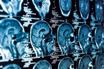
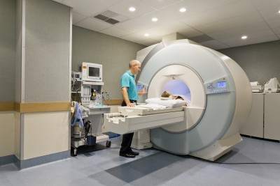
Utilizes magnets and radio waves to create detailed images of the body. Ideal for diagnosing issues related to soft tissues, discs, and the central nervous system, MRIs are more sensitive but less specific, revealing details that may not always correlate with symptoms.
CT Scan (Computed Tomography):
Combines multiple X-ray images to produce a comprehensive view of your body’s interior. Best for viewing bone injuries, diagnosing lung and chest problems, and detecting cancers. However, CT scans expose you to a higher dose of radiation compared to standard X-rays.
See Also: 4 Best Exercises For People With Sciatica
X-ray:
The quickest and most accessible form of imaging, perfect for examining fractures, infections, or abnormalities in the lungs. While X-rays are less detailed than MRIs and CT scans, they are invaluable for diagnosing specific conditions.
When to Choose Each Imaging Type
- MRI: Preferred for detailed images of soft tissues, spinal conditions, and joint problems. Essential for conditions like disc herniations, where soft tissue visualization is crucial.
- CT Scan: Optimal for quickly examining injuries or conditions that require a broader view of bone and organ systems. It’s particularly useful in emergencies.
- X-ray: Often the first step in diagnosing bone fractures or lung issues. It’s the go-to option for its speed and efficiency in revealing structural abnormalities.
Before Opting Imaging Tests, Ask Yourself Three Questions:
#1 Will the imaging pinpoint the cause of my pain?
The Paradox of MRIs and CT Scans in Diagnosing Pain
Have you ever wondered if an MRI or CT scan can pinpoint exactly where your pain originates? It’s a common question with a surprising answer: often, the answer is no. Here’s why understanding the capabilities and limitations of these powerful diagnostic tools is crucial.
The Limitations of Imaging
Misleading Results: Research shows that MRIs can reveal disc bulges and other spinal abnormalities in people without any pain—15% in 15-year-olds, 30% in 30-year-olds, and 60% in 60-year-olds. These findings challenge the assumption that visible issues on an MRI or CT scan always correlate with pain.
Sensitivity vs. Specificity: While MRIs are highly sensitive, detecting minute changes within the body, they are not always specific to the cause of pain. Dr. Christopher DiGiovanni notes that it’s rare to see a completely “normal” MRI, suggesting that abnormalities are common, even in asymptomatic individuals.
The Role of Pressure: Discs can change shape under different conditions, which means lying down during an MRI will not show the pressure that causes pain when sitting or standing. This discrepancy can lead to misinterpretations, where a patient’s symptoms don’t align with imaging results.
The Importance of Clinical Diagnosis
Beyond Imaging: A thorough history and physical examination are pivotal in making an accurate diagnosis. Sometimes, imaging findings may not reflect the patient’s experience of pain, emphasizing the need for a comprehensive evaluation by a healthcare provider.
Open MRI as a Solution: Open MRI machines, which allow for scanning in various positions, can sometimes offer a clearer picture of what happens to discs under different types of pressure. However, their availability and cost can be limiting factors.
Making Informed Decisions: MRI, CT, X-ray
Despite these challenges, MRIs and CT scans play a vital role in the diagnostic process, especially when differentiating between conditions like tumours and back pain. The key is to use them judiciously, informed by a thorough clinical assessment.
Remember, if you’re facing decisions about diagnostic imaging, discussing all aspects with your healthcare provider is crucial. Understanding the strengths and limitations of MRIs and CT scans can help you make informed choices about your healthcare journey.
#2 How will I know what is causing my pain then?
Unraveling the Mystery of Your Pain: Beyond Imaging
When you’re in pain, pinpointing the exact cause can feel like searching for a needle in a haystack. It’s natural to wonder if an MRI, CT scan, or X-ray can offer quick answers. However, the journey to understanding and addressing your pain often begins long before any images are taken.
The Central Role of Clinical Evaluation:
In-Depth Consultation: Your chiropractor or physiotherapist should start with a detailed conversation and physical examination, dedicating at least 15 to 30 minutes to understanding your symptoms, lifestyle, and any potential injury mechanisms. This initial step is crucial in forming a comprehensive view of your health status.
Diagnostic Expertise: Contrary to common belief, the true art of diagnosis lies not within the images produced by an MRI or X-ray but in the skilled interpretation of your history and physical examination findings. The overreliance on medical imaging has led to a decline in traditional diagnostic skills among some healthcare practitioners.
Formulating a Diagnosis: A proficient healthcare provider will consider your clinical presentation and may already have a primary diagnosis in mind, along with a few alternatives, based on your initial evaluation. The purpose of subsequent imaging tests like MRIs or X-rays is to confirm these suspicions rather than to discover them.
SEE ALSO: 6 Things You Should Look For In A Chiropractic Clinic: Chiropractic Clinic Best Practices
#3 Choosing the Right Diagnostic Imaging: MRI, CT Scan, or X-ray?
Deciding whether an MRI, CT scan, or X-ray is the best option for diagnosing your condition depends largely on your specific symptoms and the nature of your ailment. Understanding the strengths and applications of each imaging technique can guide you and your healthcare provider toward the most effective diagnostic approach.
When to Consider Each Imaging Option: MRI, CT Scan or X-ray
X-ray for Osteoarthritis:
- Ideal for Initial Diagnosis: X-rays are often sufficient for diagnosing osteoarthritis, characterized by morning stiffness that improves after movement.
- Cost-effective: Compared to MRI and CT scans, X-rays are more affordable and widely accessible for assessing joint wear and tear.
MRI for Disc Herniations:
- Superior Soft Tissue Imaging: MRIs excel in detailing soft tissue conditions, making them perfect for identifying disc herniations.
- Non-invasive Diagnosis: Before opting for an MRI, consider chiropractic care, extension exercises or Yoga Cobra, which can effectively treat many cases of disc herniation.
See Also: The Best Exercises For Your Herniated Disc
MRI for ACL Tears:
- High Sensitivity: MRIs provide detailed images of soft tissues, including ligaments, making them more sensitive for detecting ACL tears than physical examinations alone.
- Comprehensive Damage Assessment: An MRI can also reveal associated cartilage and ligament injuries, offering a complete picture of the knee’s condition.
CT Scan for Fractures Invisible on X-rays:
- Enhanced Bone Imaging: CT scans are superior for visualizing bone fractures that X-rays may miss, such as rib fractures.
- Critical for Complex Diagnoses: In cases where a detailed bone structure view is necessary, CT scans offer the clarity needed for accurate diagnosis.
Arthrograms for Cartilage Tears:
- Specialized Imaging: Injecting dye into a joint before taking an X-ray, MRI, or CT scan can highlight cartilage tears more clearly, essential for diagnosing meniscus, hip labral, and shoulder labral tears.
Key Considerations When Choosing Between An MRI, CT and X-ray
- Symptom-Specific Imaging: The choice between MRI, CT scan, and X-ray should be guided by the specific symptoms and the area of the body affected.
- Consult with Your Healthcare Provider: A thorough clinical evaluation is crucial in determining the most appropriate imaging technique for your condition.
- Cost vs. Benefit: Consider the cost and diagnostic value of each imaging option, especially when dealing with conditions that might be managed with less expensive alternatives.
Dangers of an MRI
An MRI machine has a powerful magnet that attracts most metallic objects. Therefore the danger lies with any implants that contain iron. Your implant can move and injure you physically or heat up and even burn you. The following are implants that are unsafe for MRIs.
- Older vascular stents
- Cochlear implants
- Brain-aneurysm clips
- Pacemakers or cardiac defibrillators
- Gastrointestinal cips
- Certain medication pumps (such as insulin pumps)
- Stainless steel spinal fusion screws and plates cause problems for the MRI and you. However, titanium screws and plates are fine. The stainless steel causes the digital picture to be degraded.
- Metallic objects like metal filings that have accidentally gone into the eye.
Gadolinium dyes are used in some MRIs, Gadolinium contrast dye is safe for most of you, but unsafe and can cause death in those with bad kidneys. If you are getting a procedure with Gadolinium your medical doctor should do a screening test for your kidney function.
Understanding the Radiation Risks: CT Scans, X-rays, and the Safety of MRIs
When it comes to medical imaging, understanding the risks and benefits of each option is crucial. While CT scans and X-rays are invaluable tools in diagnosing various conditions, it’s important to be aware of the radiation exposure involved. MRI scans, on the other hand, offer a radiation-free alternative for many diagnostic needs. Let’s dive deeper into the implications of choosing between these imaging techniques.
Radiation Exposure: A Comparative Look
Everyday Radiation vs. Medical Imaging
- On average, we’re exposed to about 2 millisieverts (mSv) of radiation annually from natural sources like rocks, the sun, and even aeroplane flights.
- Residents in high-altitude areas, such as Colorado, receive around 10 mSv due to less atmospheric protection.
CT Scans and X-rays: How Much Radiation Do You Get?
- A neck CT scan exposes you to about 6 mSv, roughly three years’ worth of natural radiation.
- A brain CT scan equals about 2 mSv, or one year’s worth of background radiation.
- A low back X-ray comes in at 1.5 mSv, about eight months’ worth of natural exposure.
- Neck X-rays and mammograms offer lower exposures, at 0.2 mSv and 0.8 mSv respectively, but these still add up over time.
Assessing the Need for Imaging
Before opting for a CT scan or X-ray, consider the following:
- Diagnosis Clarity: If your diagnosis is uncertain, imaging may be crucial.
- Treatment Impact: Will the scan results change your treatment approach? If not, reassess its necessity.
- Previous Scans: Remember, radiation exposure is cumulative. Evaluate the necessity of additional CT scans carefully.
- Cancer Risk: Those at high risk or with a family history of cancer should weigh the benefits and risks more carefully.
- Pain Localization: Can the scan accurately identify the source of your pain?
MRI: A Safer Alternative?
MRIs stand out as they do not use ionizing radiation, making them a safer choice in many scenarios. Despite advances in medical technology, it’s essential to consider the historical context—lessons learned from past practices, like the widespread use of X-rays in pregnant women during the 1960s, underscore the importance of caution and informed decision-making in medical imaging.
Making an Informed Choice: MRI, CAT scan and X-ray
Choosing between an MRI, CT scan, or X-ray involves a careful evaluation of your specific situation, guided by the expertise of your healthcare provider. By understanding the strengths and limitations of each imaging method, you can make informed decisions about your health care.
Tell us what you think in the comments below and like us on Facebook. This Toronto Downtown Chiropractor will answer all questions in the comments section.


I am a 50 year old female. Over three years ago, I lifted 10 pound hand weights above my head and began getting pain around my right breast in the rib area. I have been to a rheumatologist and he did ct scan and X-ray and nothing was found wrong. Have had heart checked, and full check by gastrointestinal doctor. All shows up clear including blood work all normal! I do have tenderness when touching area around right breast. It also hurts to twist torso and stretch the right side at times! What do you think?
Author
Thanks for your question Kristi. Sounds like you strained a muscle or possibly the rib. While I am glad you ruled out the more serious conditions I would have personally gone to a chiropractor or physiotherapist first as you did something mechanical (lifting up a weight). So likely it will be a mechanical problem. You should always check that out first with someone that can examine you.
That’s my opinion and not my recommendation. I will do my best to answer your questions to help you as much as possible. Hope that helps.